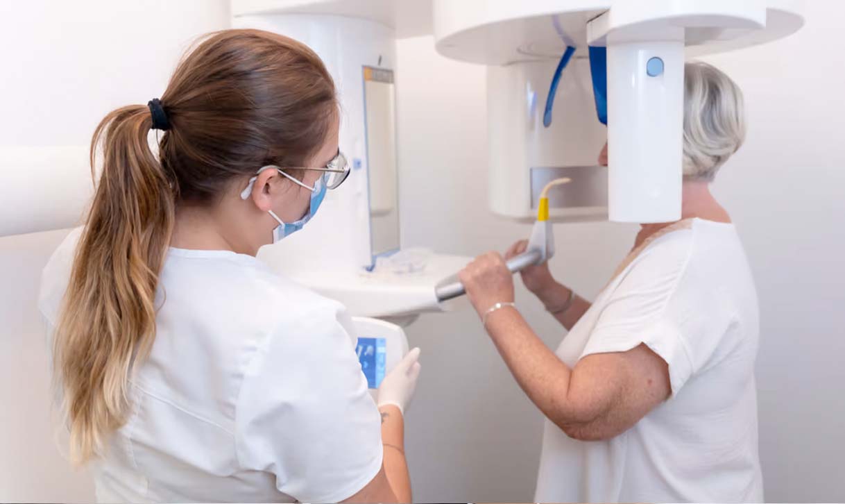
Tomography and Panoramic Imaging
Dental tomography is a radiological diagnostic method used to create a cross-sectional image of the area to be examined using x-ray. With the tomography image, bone and soft tissue details that are not seen in normal x-rays can be seen. In our center, Morita brand Veraviewepocs 3D device, which works with the cone beam principle, is used.
With the dental tomography device, many sections are taken from axial sagittal and coronal planes. These sections are then re-sliced and restructured using advanced software so that the targeted area can be viewed from any angle and from any direction. These sections, which provide high diagnostic detail and measurement accuracy, serve as a guide in all kinds of treatment. Additionally, the bone structure can be examined by creating a volumetric image of the skull.
DENTO MAXILLO FACIAL SURGERY
Empacted and displaced teeth (wisdom teeth are extra teeth).
Apical periodontitis jaw cysts.
Pre- and postoperative imaging of important anatomical markers and structures.
Cleft palate patients.
Trauma cases (bone and tooth fractures).
Visualization of joint TMJ.
IMPLANTOLOGY
Implant planning and retrieval
Detection of bone thickness and tissue status
Determination of distances to the mandibular canal or sinus floor
PERIODONTOLOGY
Implant planning and retrieval
Detection of bone thickness and tissue status
Determination of distances to the mandibular canal or sinus floor
ENDODONTIC
Configuration and appearance of root canals.
Root canal measurements.
Detection of anatomical deviations and unusually large root canals.
ORTHODONTIC
Shaping the roots.
Anatomical situation, relationship between teeth.
Periodontal ligament (ankylosis).
Resorptions


